

In recent years, there has been a significant advancement in the field of Artificial Intelligence (AI) and Augmented Reality (AR). These technologies have become increasingly popular and have the potential to enhance virtual experiences in various fields such as gaming, education, healthcare, and...
How a Digital Pathologist Determines Why a Plant Died
92
18.08.2026
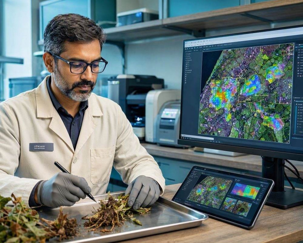
A dead plant often reveals only vague external symptoms: wilted leaves, discolored stems, weak growth, or collapsed branches. These signs indicate failure but rarely identify its origin. A digital pathologist can investigate the cause by examining a high-resolution image of a stem, root, or branch cross-section. Tissue structure, color, lesion shape, and vascular condition provide evidence that may distinguish infection from drought, mechanical injury, chemical damage, or natural decay.
Why a Cross-Section Is So Informative
Plants transport water through xylem and distribute sugars through phloem. When these tissues are blocked, destroyed, or separated, the organs above the damaged area may die. A clean cross-section exposes the arrangement of vascular bundles, annual growth rings, bark, pith, and surrounding tissues. Abnormalities that remain hidden beneath the surface become measurable features in a digital image.
Vascular Discoloration
Brown, black, or reddish streaks within the xylem may indicate vascular wilt, bacterial infection, or prolonged oxidation after tissue death. Their distribution matters. A continuous dark ring suggests a different process from isolated spots or discoloration restricted to one side. Digital analysis can map these regions and calculate how much of the water-conducting tissue has been affected.
Rot, Cavities, and Tissue Collapse
Softened areas, hollow zones, irregular cracks, and loss of cellular structure can point to fungal decay or secondary colonization. However, rot observed after death is not automatically the primary cause. The pathologist must determine whether decomposition began while the plant was alive or developed later in already damaged tissue.
How Digital Diagnosis Works
A reliable assessment combines image processing with botanical interpretation. The digital pathologist does not merely match colors. It evaluates patterns, tissue boundaries, anatomical location, and supporting information about the plant’s environment.
- Image preparation: The system checks focus, lighting, scale, cutting artifacts, and color accuracy before interpreting biological features.
- Tissue segmentation: Computer vision separates bark, cambium, xylem, pith, lesions, cavities, and visibly discolored regions.
- Pattern comparison: Detected abnormalities are compared with reference images representing diseases, water stress, frost injury, and physical damage.
- Probability ranking: The system produces a differential diagnosis rather than claiming certainty from one visual feature.
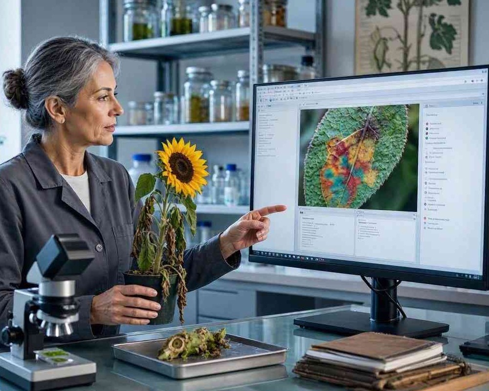
Clues That Strengthen the Conclusion
Image findings become more useful when paired with the plant species, age, recent weather, irrigation history, soil condition, and progression of symptoms. For example, blocked vessels combined with sudden wilting and a known susceptible host support vascular disease. Narrow growth rings and intact vessels may instead favor chronic drought or root restriction.
Common Causes Visible in Cross-Sections
Different forms of plant failure can produce recognizable internal patterns. Although microscopy or laboratory testing may still be required, a cross-section helps narrow the investigation quickly.
- Vascular pathogens may create dark streaks or sector-shaped lesions.
- Root suffocation can cause diffuse browning and poorly developed tissue.
- Frost may produce sharp cracks and localized cambial death.
- Mechanical injury often affects one side and disrupts normal growth rings.
- Wood-decay fungi may leave cavities, pale zones, or fibrous tissue.
Why Human Review Still Matters
Similar patterns can have different causes, and image quality can distort color or texture. A cut made with a blunt tool may resemble tissue collapse, while staining from storage may imitate disease. For that reason, digital pathology should support an expert rather than replace one. A plant pathologist can request microscopy, fungal culture, molecular testing, or soil analysis when visual evidence is inconclusive.
From Image Evidence to Practical Action
The final report should identify the most likely cause, explain the visible evidence, state the confidence level, and recommend confirmation steps. It may also advise removing infected material, disinfecting tools, improving drainage, or testing nearby plants. By turning hidden anatomical changes into documented evidence, a digital pathologist makes plant-death investigations faster, more consistent, and easier to verify.